JUNIORBONES
Orthopaedics and trauma for junior orthopaedic trainees and medical students
  • Home
    • About
  • Patient evaluation
    • History
    • Examination >
      • Shoulder examination
      • Hip examination
      • Knee examination
      • Foot & ankle examination
      • Spine examination
  • Trauma
    • Orthopaedic emergencies
    • Common trauma referrals >
      • Proximal humerus injuries >
        • Shoulder dislocation
        • Proximal humerus (shoulder) fractures
      • Upper-limb long bone fractures
      • Elbow injuries >
        • Adult elbow fractures
        • Paediatric supracondylar elbow fracture
      • Distal radius fractures
      • Spine trauma
      • Pelvic & acetabular fractures
      • Proximal femur fracture
      • Lower-limb long bone fractures
      • Knee injuries >
        • Fractures at the knee
        • Soft tissue knee injuries
      • Foot & ankle injuries >
        • Ankle fractures
        • Foot fractures
      • Peri-prosthetic fractures
      • Paediatric trauma
      • Septic arthritis >
        • Septic native joints
        • Septic joint replacement
  • Orthopaedic conditions
    • Osteoarthritis
    • Inflammatory & infective arthritis
    • Other joint disorders
    • Spine
    • Paediatric orthopaedics
    • Orthopaedic pathology
  • Cases blog
  • Trauma guidelines
  • Book downloads
  • Authors
  • Contact

What is Gamekeeper's Thumb?

28/6/2015

0 Comments

 
Melissa a 26 year old female, RHD, presented to her local GP with ongoing pain and swelling on the ulnar aspect of the base of her right thumb. 2 weeks ago, whilst on a 1 month cycling trip around Europe she fell off her bike and her thumb was forcefully abducted, caught on the bike handle. She had immediate pain but continued her tour. This is the first management she has sought for her thumb. 

What is Gamekeepers Thumb?
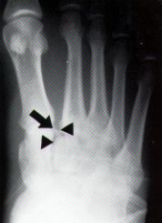
Avulsion or rupture of the ulnar collateral ligament (UCL) - see image below


Mechanism of injury: hyper abduction or extension of MCP joint of thumb

Epidemiology: gamekeepers repetitively stressed the UCL ligament and MCP joint of their thumb when breaking rabbits necks (chronic injury). Today more commonly seen in skiers, also known as 'Skiers Thumb,' occurs when the stock or stock strap forcefully abduct the skiers thumb when falling or aggressively planting the pole (acute injury). 

Stener Lesion: UCL ligament avulsed above the adductor aponeurosis. Adductor aponeurosis and adductor pollicis muscle now lying between ligament and proximal phalanx, will not heal without surgical repair.
Picture
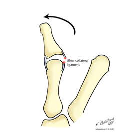
Ruptured UCL: Gamekeeper's Thumb. Gaillard, F (2009)
Picture
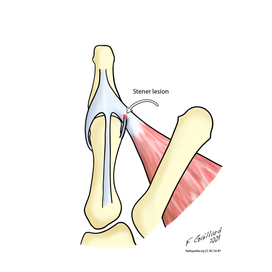
Stener Lesion: UCL avulsed and retracted above adductor aponeurosis and adductor pollicis muscle. Gailard, F (2009)
Physical Examination specific to UCL ligament rupture:
Appearance and palpation - mass from the torn UCL ligament or bony avulsion may be present on the ulnar aspect of the 1st MCP
Stress MCP joint radially: 
  1. Neutral position: if lax indicative of accessory UCL injury
  2. 30 degrees of flexion: if lax indicative of proper UCL injury
  3. ALWAYS COMPARE TO OTHER SIDE
valgus laxity in both flexion and extension = complete UCL rupture
Picture
What imaging would you order?
1. X-rays: AP, lateral, oblique - can show bony avulsion
If there is no bony avulsion, look for ulnar side of the MCP joint, if it appears widened this is suggestive of UCL injury. Stress views were previously undertaken in this situation, however, now there is believed risk of worsening the injury and creating a Stener lesion.

2. Ultrasound scan: to identify tear and Stener lesion, however, operator dependent. 

3. MRI: gold standard in identifying; discontinuity of ligament and or joint capsule, boney oedema and Stener lesion. 

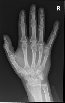
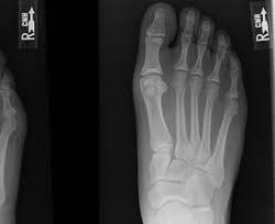
Describe Mel's AP X-ray (image to left):
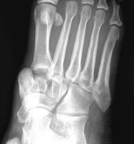
Always ensure you identify patient and search for old X-rays for comparison, and all views available
There is a minimally displaced intra-articular avulsion fracture of the ulnar aspect of the proximal phalanx of the right thumb.
Always ensure you look for additional fractures, soft tissue swellings, etc. Remember here could be more than one injury.


Treatment Options: Non Operative vs Operative 
Non-operative: Immobilise in cast for 4-6 weeks
Indication: partial tears (<20 degrees of side to side variation). (McKean, J. 2014)

Operative:
1. Ligament Repair: using sutures, anchors, screws
2. Ligament Reconstruction: tendon graft
3. MCP Fusion: for chronic injury and pain, often if the above methods have failed 
Indication:
  1. Acute injury with >20 degrees of varus/valgus instability 
  2. >35 degrees of opening
  3. Stener lesion: as described and depicted above

References: 
Dawes, L., Weerakkody, Y et al. Gamekeepers Thumb. Radiopaedia. Retrieved 27 June 2015 from http://radiopaedia.org/articles/gamekeeper-thumb

Gaillard, F. (2009). Gamekeepers Thumb. Radiopaedia. Retrieved 27 June 2015 from http://radiopaedia.org/articles/gamekeeper-thumb

McKean, J. (2014) Thumb Collateral Ligament Injuries. Orthobullets. Retrieved 27 June 2015 from http://www.orthobullets.com/hand/6040/thumb-collateral-ligament-injury

0 Comments

Lisfranc Injury

23/6/2015

1 Comment

 
24 y.o male has been referred to fracture clinic query a right foot, Lisfranc injury, He presented to ED 2 days ago with inability to weightbear, forefoot pain and swelling after a tackle in a rugby game.

What is the Lisfranc ligament and why is it important?
It is a ligament (depicted above in yellow) from medial cuneiform (under 1st metatarsal) to the base of the 2nd metatarsal on the plantar surface. It tightens with abduction and pronation of the forefoot.
It is integral in maintaining the midfoot arch and stabalising the 2nd metatarsal. As there is no ligament directly connecting the 1st and 2nd metatarsal. 

What is the mechanism of injury usually?
Injury usually includes fall from height, motor vehicle accident or athletic injury like in this case.

Hyperplantar flexed forefoot, compression (axial load) and abduction (indirect rotational force) transmitted to the tarso-metotarsal articulation – causing metatarsals to be displace in a dorsal/lateral direction, as depicted below.
[1] Bloomberg J (2015).
Assessing the patient:
1. Assess foot circulation
  • The anterior tibial artery has the first dorsal and first plantar metatarsal artery that supplies the medial foot, disruption of this astemosis can cause signficiant haemorrhage and compartment syndrome
  • Compartment syndrome

2. Assess soft tissue
  • Stellate bruising over midfoot plantar area
  • Swelling, prominence of medial tarsal bones, shortening of forefoot

3. Assess associated injury

  • Abduction mechanism of injury – compression fracture of cuboid, MTPJ dislocations, TMTJ fractures/dislocations, metatarsal fractures

Instability test: grasp metatarsal heads and apply dorsal force to forefoot while other hand palpates the TMT joints dorsal subluxation suggests instability
  • If 1st and 2nd  metatarsals can be displaced medially and laterally, global instability is present and surgery is required
  • No dorsal subluxation = plantar ligaments are intact, injury may be treated non-operatively

Provocative tests may reproduce pain with pronation and abduction of forefoot


What imaging would you order?

Xrays: AP, lateral, oblique 30 degrees, If possible -AP Weight bearing Xrays: comparison can be made to other side
Stress radiograph: if non-weight bearing Xrays normal but clinically high suspicion for Lisfranc stress views may be helpful to show instability
CT scan: for diagnosis and preoperative planning
MRI: diagnose pure ligamentous injury

 
Below are his Xrays, describe the deformity.
Picture
AP [2]
Picture
Lateral [2]
Picture
Oblique [2]
Picture
Flec Sign [1]
What are the 5 critical radiographic signs of midfoot instability?
AP view:
1. The medial base of the 2nd metatarsal does not align with the medial side of the middle cuneiform.
2. Widening of the interval between the first and second ray

Oblique view:
3. Medial side of  4th metatarsal base does not line up with medial side of cuboid

Lateral view:
4. Dorsal displacement of metatarsal base
5. Disruption of the medial column line (line tangential to the medial aspect of the navicular and the medial cuneiform)

Flec sign (on AP view): may see bony fragment  in 1st intermetatarsal space (avulsion of Lisfanc ligament from base of 2nd metatarsal) 

Treatment
Non-operative: immobilization for 8 weeks Vs Operative

Indications for non-operative management?
Non-displaced on weight-bearing and stress radiographs and no evidence of bony injury on CT. OR:
  • Non-ambulatory patients
  • Severe vascular disease
  • Severe peripheral neuropathy
  • Instability in only the transverse plane

What are the different operative options:
1.    Open reduction and rigid internal fixation:
Indication: Any instability (> 2mm shift) and or fracture dislocations, rather than pure ligamentous injuries

2.    Primary athrodesis of the 1st, 2nd and 3rd metatarsal joints
Indication: pure ligamentous injury

3.    Midfoot athrodesis
Indication: unstable midfoot architecture with progressive arch collapse and forefoot abduction, chronic lisfranc injuries that have led to midfoot arthrosis and failed conservative management

Reference:
[1] Blomberg, J (2015)  Lisfranc Injury (Tarsometatarsal fracture-dislocation). Orthobullets. Retrieved 20 June 2015 from http://www.orthobullets.com
[2] And Rabou, A., Gaillard, F et al. (2015) Lisfranc Injury. Radiopaeidia. Retrieved 20 June 2014 from http://radiopaedia.org/articles/lisfranc-injury


1 Comment

Anterior Shoulder Dislocation

7/6/2015

0 Comments

 
Picture
[1] Cunningham, N. Fennessey, G. (2012)
Picture[2]
Read the case below and attempt to answer the questions. 
Steven, a 19 year old professional basketball player, presents to the ED  with right shoulder pain and deformity after his arm was pulled backwards when he was defending another player in a basketball game 2 hours ago.

1. From the photo of Steven to the left, describe what you see?
There is obvious asymmetry between Steven’s shoulders, his right acromion and humeral head are prominent, his right arm is slightly abducted and internally rotated, and is being supported by his left hand.

2. Describe the likely mechanism of injury?
The shoulder is abducted and externally rotated when an anterior force is applied to the arm.

3. What focused physical examination would you undertake?
ALWAYS – check neurovascular status, rule out concurrent injuries (brachial plexus, vascular and cervicle)
5% of anterior dislocations are associated with axillary neuropraxia- should resolve within weeks. It will be difficult to assess deltoid function, therefore check dermatomal innervation (skin over distal deltoid) AND remember the entire brachial plexus is also at risk, not just the axillary nerve.

Apprehension Sign: patient is supine with shoulder off side of bed, abduct shoulder to 90 degrees and elbow to 90 degrees then externally rotate shoulder, positive test is facial expression of ‘apprehension’

Relocation Sign: decreased apprehension with anterior force applied on shoulder, when conducted apprehension maneuver descrived above.

Sulcus Sign: arm adducted by side, place inferior traction on elbow, if a depression occurs just below the acromion, then sulcus sign is positive.


4. What imaging would you order?
1. Xray – AP, lateral (scapular Y), axillary (if possible)
            Other views: stryker view (shows Hill-Sacks lesion), west point view (shows glenoid bone loss)

2. CT if delayed presentation, complex fracture dislocation, osteoperotic patient


5. An AP Xray was taken of Steven's right shoulder, describe it and the diagnosis.

Always start with: Patient details, date, any previous Xrays to compare to, any additional views.
AP view: Head of humerus is anterior to the glenoid and inferior to the coracoid.
Always remember to ask for lateral/other views?
Diagnosis: subcoracoid anterior dislocation.

6. What closed reduction methods could you use to relocate Steven's shoulder?
Closed Reduction manoeuvres
Ensure adequate analgesia, ideally anaesthetic involvement.
1.   Cunninghams Method: Ensure patient sits up straight during the entire maneuver. Have patients arm adducted, elbow at 90 degrees and arm in neutral position. Begin by firmly massaging trapezius muscle on affected side, beginning proximally and working towards shoulder, then deltoid and finally massage biceps muscle
2.   Kocher’s tractionless manoeuvre: Humerus adducted, elbow flexed at 90 degrees and arm in neutral position. Then externally rotate arm and humerus (keeping humerus adducted to body) then move humerus anteriorly (keeping body still) and finally internally rotate arm.

7. You have successfully reduced Steven's shoulder, what is the next step?
1.     Place in neutral brace
2.     Recheck neurovascular status
3.     Repeat Xrays
4.     Refer to fracture clinic within 1/52

8. What injuries are associated with anterior dislocations?

1.     Labral and cartilage injuries:
  1. Bankart Lesion: avulsion of the anterior labrum and anterior band of the IGHL – present in 80% of patients with anterior dislocations
  2. Humeral avulsion of the glenohumeral ligament
  3. Glenoid labral articular defect
  4. Anterior labral periosteal sleeve avulsion


2.     Fractures:
  1. Hill-Sachs deformity: a compression fracture of the posterosuperior aspect of the humeral head 
  2. Bony Bankart lesion: fracture of the anterior inferior glenoid
  3. Greater tuberosity fracture
  4. Lesser tuberosity fracture


References:
[1] Cunningham, N., Fennessey, G. (2012) Shoulderdislocation.net. Retrieved on 6 June 2015 from http://shoulderdislocation.net/anatomy/dislocated-anatomy
[2] Matthew, S., Stevenson, H. Physio-pedia. Retrieved on 6 June 2014 from http://www.physio-pedia.com/Shoulder_Dislocation#References
0 Comments

    AuthorAc

    Francois Tudor and James Donaldson recently completed their orthopaedic training and wish to share their knowledge and interest in orthopaedics. This blog will provide weekly interesting cases that we hope will help you learn and develop your knowledge in orthopaedics, updated by resident, Moni Brunt-Mackenzie 

    Archives

    December 2015
    October 2015
    September 2015
    August 2015
    July 2015
    June 2015
    March 2015
    January 2015
    November 2014
    October 2014

    Categories

    All

    RSS Feed

Powered by Create your own unique website with customizable templates.