Assessment and management of supracondylar fractures in children
by Renae Vardi
SCENARIO: You are called to the Emergency Department to assess a 5 yo who hurt her right elbow falling from a swing.
HISTORY:
Age
Hand dominance
Mechanism of injury - include where and when, who saw it happen
Time last ate and drank
Past medical & surgical history
Medications
Allergies
EXCLUDE NON-ACCIDENTAL INJURY (if concerns, involve local paediatricians)
With a fall onto an outstretched hand - consider supracondylar fracture or fractureof the lateral condyle of the elbow. Also consider radial head fracture, elbow dislocation, Monteggia fracture or distal radius/ulna fracture.
Direct blow on to Flexed elbow - consider flexion type supracondylar fracture, radial head dislocation, radial head/neck fracture
Immediate assessment:
Exclude all other injuries
Examine affected limb - look for obvious deformity and determine if open or closed injury.
Examine radial/ulna/median nerves both motor and sensation (difficult in young child. Ask them to give you thumbs up and give the "OK" sign with fingers). Palpate for a radial & ulna pulse. Assess capillary refill.
Investigations:
X-Ray of affected limb - true AP and lateral and images of joint above and below.
|
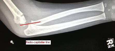
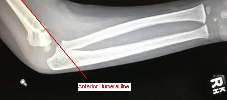
Radiological lines to consider:
1. Radio-capitellar line: Line drawn down neck of radius on AP film - should pass through the center of the capitellum) Used to assess for radial dislocation. 2. Anterior humeral line: A line drawn on a lateral view along the anterior surface of the humerus should pass through the middle third of the capitellum. In a displaced supracondylar fracture, this line will lie in front of the capitellum. |
Gartland Classification
Type I - Nondisplaced - anterior humeral line intersects the capitellum, an intact olecranon fossa, no medial or lateral displacement.
Type II - Partially displaced - usually fragment is extended but not completely translated with some cortical contact. The anterior humeral line does not intersect the capitellum. Some rotational displacement and tilt into varus may be present. Subdivision:
IIa - minimally displaced, no rotation
IIb - rotational deformity
Type III - Displaced - circumferential break in the cortex with displacement of the fracture fragments. The distal fragment is generally displaced posteriorly with the metaphyseal fragment impaled into the brachialis muscle and anterior soft tissues.
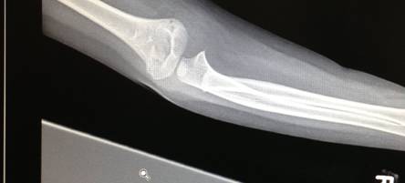
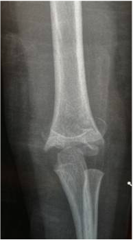
Anterior-posterior X-ray of a Gartland I fracture. Note no displacement of fracture line.
|
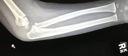
X-rays of a Gartland I fracture. Note extension of distal fragment but no displacement. Note anterior humeral line broken.
|
Anterior-Posterior X-ray of a Gartland III fracture. Note overlap of fragments and shortening.
|
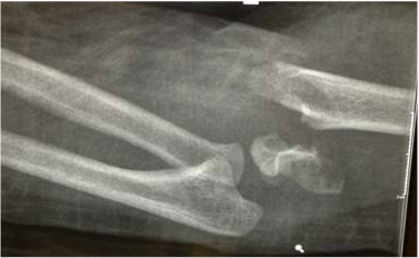
Lateral X-ray of a Gartland III fracture. Note posterior displacement, shortening and angulation. Be aware of vascular or nerve injuries in this type.
|
Management
Dictated by grade of injury:
Grade I - collar and cuff, elbow flexed to 130degress. May require admission for vascular obs overnight. Review in fracture clinic 1/52 XROA
Grade IIA - as per grade I
Grade IIB - Manipulation Under Anaesthetic +/- k-wires
Grade III - Manipulation Under Anaesthetic & probably k-wires
ALWAYS ASSESS PULSES
A pulseless supracondylar is an orthopaedic emergency. Arrange transfer to operating theatre ASAP and get urgent senior review.
Consider compartment syndrome if very painful or very swollen, especially in displaced fracture.
Pulses may return with reduction under anaesthetic or may require exploration with vascular/plastic surgeons +/- fasciotomy.
Complications
Early: Anterior Interosseous Nerve or other nerve plasy, Brachial artery injury, compartment syndrome
Late: Cubitus valgus deformity (due to malunion), flexion contracture (often due to missed compartment syndrome)
Late: Cubitus valgus deformity (due to malunion), flexion contracture (often due to missed compartment syndrome)
