JUNIORBONES
Orthopaedics and trauma for junior orthopaedic trainees and medical students
  • Home
    • About
  • Patient evaluation
    • History
    • Examination >
      • Shoulder examination
      • Hip examination
      • Knee examination
      • Foot & ankle examination
      • Spine examination
  • Trauma
    • Orthopaedic emergencies
    • Common trauma referrals >
      • Proximal humerus injuries >
        • Shoulder dislocation
        • Proximal humerus (shoulder) fractures
      • Upper-limb long bone fractures
      • Elbow injuries >
        • Adult elbow fractures
        • Paediatric supracondylar elbow fracture
      • Distal radius fractures
      • Spine trauma
      • Pelvic & acetabular fractures
      • Proximal femur fracture
      • Lower-limb long bone fractures
      • Knee injuries >
        • Fractures at the knee
        • Soft tissue knee injuries
      • Foot & ankle injuries >
        • Ankle fractures
        • Foot fractures
      • Peri-prosthetic fractures
      • Paediatric trauma
      • Septic arthritis >
        • Septic native joints
        • Septic joint replacement
  • Orthopaedic conditions
    • Osteoarthritis
    • Inflammatory & infective arthritis
    • Other joint disorders
    • Spine
    • Paediatric orthopaedics
    • Orthopaedic pathology
  • Cases blog
  • Trauma guidelines
  • Book downloads
  • Authors
  • Contact

Assessment and management of proximal humerus dislocations
by Toby Baring

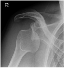
Figure 2: Posterior dislocation of the shoulder - internal rotation of the head looks gives it a symmetrical appearance also known as the "light bulb" sign
History/mechanism
Anterior
  • Typically hyper-external rotation of the shoulder when fully abducted (dislocation can occur without abduction)
  • Seizure or electric shock
  • Holding arm, possibly in neutral rotation, prominant acromion as deltoid is stretched over it


Posterior
  • Axial loading of arm with internal rotation and flexion of the shoulder
  • Seizure or electric shock
  • Arm held against body(internally rotated), may complain of “shoulder stiffness” only

Assessment
Full neurovascular assessment of the effected arm
Rule out concurrent injuries (e.g. brachial plexus/vascular/cervical)

Investigations
To determine direction of dislocation and extent of bony injury
  • X-ray – two orthogonal views
If patient known to have recurrent shoulder dislocations and a low energy injury then reduction maneuver can be attempted without x-ray
  • CT scan if delayed presentation or complex fracture dislocation
Anterior shoulder dislocation
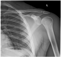
AP x-ray demonstrating anterior dislocation of the shoulder - typically the head sits inferior to the glenoid
posterior shoulder dislocation
AP x-ray demonstrating posterior dislocation of the shoulder - internal rotation of the head looks gives it a symmetrical appearance also known as the "light bulb" sign
Management
  • If simple dislocation or isloated avulsion of greater tuberosity (GT), attempt reduction with analgesia and sedation (ideally by anaesthetics)
  • If complex fracture dislocation make arrangements for patient to go to next available theatre session during daylight hours with upper limb specialist. Consent for closed reduction +/- open reduction +/-  fracture fixation +/-hemiarthroplasty.
Reduction maneuvers
Anterior dislocation
  • Kocher’s tractionless maneuver
  • Hippocratic technique
Posterior dislocation
  • Abduction and external rotation
  • Hippocratic  technique
If reduction successful
  • Place in a neutral rotation brace ideally (particularly if a fracture of the GT has anatomically reduced)
  • Posterior dislocation - neutral rotation brace is obligatory (simple sling may cause re-dislocation)
  • Re-assess neurovascular status of arm
  • Repeat x-ray – two orthogonal views
  • Refer to upper limb fracture clinic – to be seen within 1 week

If reduction unsuccessful
  • Make arrangements for patient to go to next available theatre session during daylight hours. Consent for closed +/- open reduction
  • In the case of a fractured GT if the shoulder reduces but the GT remains >5mm displaced discuss with an upper limb specialist within 48hrs
Further comments/pitfalls/exceptions
  • Careful eliciting the history, i.e. position of the arm, should allow one to diagnose the direction of dislocation (see table).
  • Recurrence dislocations may occur with ease (turning over in bed, reaching out to grab an object) and should be treated as per a primary dislocation in the acute phase (i.e. reduction and sling).
  • Examination of the neurology should concentrate on assessment of the axillary nerve – deltoid function is difficult to assess with a dislocated shoulder but the sensation of the skin of the regimental badge area will give an indication of the nerve’s function. NB – the axillary is not the only at risk nerve from a shoulder dislocation so please assess the other upper limb nerves with care.
  • In polytrauma patients, diagnosis and treatment of the shoulder dislocation is part of the secondary survey. Make sure the primary survey is complete and the patient is stable prior to attempting reduction of a shoulder dislocation.
  • Isolated greater tuberosity fracture dislocations in osteoporotic bone – these may well be associated with occult fractures of the anatomical neck or lesser tuberosity. Any patient at risk of osteoporosis should have a CT prior to any reduction maneuver. If a complex fracture is diagnosed then the patient should undergo open reduction to minimize the risk of fracture displacement.
  • Nerve injuries are usually transient and should be only monitored in the initial instance.
  • Habitual dislocators – these are typically young females who voluntarily dislocate their shoulder for attention seeking purposes (often as part of a psychological disorder). These patients may been well known to the A+E department. Discuss with your senior colleague prior to attempting dislocation.
  • Luxatio erecta – this is pure inferior dislocation of the shoulder (very rare). The patient will present with the arm in fixed abduction or held behind their head. This condition has a high risk of neurovascular injury. Discuss with senior prior to attempted reduction.
shoulder dislocation luxatio erecta
AP x-ray shoulder demonstrating luxatio erecta - pure inferior subluxation of the glenohumeral joint with the arm abducted
shoulder fracture dislocation
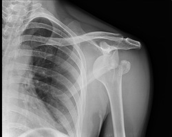
AP x-ray of shoulder demonstrating anterior dislocation with GT fracture
Powered by Create your own unique website with customizable templates.