JUNIORBONES
Orthopaedics and trauma for junior orthopaedic trainees and medical students
  • Home
    • About
  • Patient evaluation
    • History
    • Examination >
      • Shoulder examination
      • Hip examination
      • Knee examination
      • Foot & ankle examination
      • Spine examination
  • Trauma
    • Orthopaedic emergencies
    • Common trauma referrals >
      • Proximal humerus injuries >
        • Shoulder dislocation
        • Proximal humerus (shoulder) fractures
      • Upper-limb long bone fractures
      • Elbow injuries >
        • Adult elbow fractures
        • Paediatric supracondylar elbow fracture
      • Distal radius fractures
      • Spine trauma
      • Pelvic & acetabular fractures
      • Proximal femur fracture
      • Lower-limb long bone fractures
      • Knee injuries >
        • Fractures at the knee
        • Soft tissue knee injuries
      • Foot & ankle injuries >
        • Ankle fractures
        • Foot fractures
      • Peri-prosthetic fractures
      • Paediatric trauma
      • Septic arthritis >
        • Septic native joints
        • Septic joint replacement
  • Orthopaedic conditions
    • Osteoarthritis
    • Inflammatory & infective arthritis
    • Other joint disorders
    • Spine
    • Paediatric orthopaedics
    • Orthopaedic pathology
  • Cases blog
  • Trauma guidelines
  • Book downloads
  • Authors
  • Contact

Assessment and management of upper limb long bone fractures
by James Donaldson

Humeral shaft fracture

History:

Age – bimodal age distribution: young high energy injury or elderly osteoporotic
Occupation and handedness – useful for deciding treatment, rehab protocol, compliance
Time/date of injury

Past medical/surgical history – including malignancies past and present
Medication/drugs and allergies
Last ate/drank (for timing of emergency surgery if needed)

Radiographs:
  • AP and lateral of humerus including the joint above and below.

humerus fracture
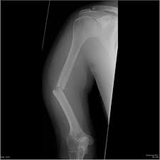
AP x-ray of humerus shaft fracture
Symptoms and Signs
  • Pain, weakness
  • Deformity, swelling
  • Assess skin status if sharp bony spikes present
  • Assess for other injuries
  • Neurovascular exam is critical
            - Especially radial nerve status

Discuss with senior urgently if open or neurovascular injury
Initial Treatment
  • Analgesia
  • Collar and cuff or coaptation splint
  • Majority can be treated non-operatively
  • Acceptable alignment for non-operative treatment
            - <20-30 degrees varus or valgus angulation
            - <20 degrees anterior angulation
            - <3cm shortening
Definitive Treatment
  • Once the swelling has settled (around 1 week) a functional humeral brace can be applied
  • Possible indications for surgery (ORIF or IM nail):
            - Open fracture
            - Vascular compromise
            - Brachial plexus injury
            - Ipsilateral forearm #
            - Bilateral humeral #s
            - Pathological #
            - Polytrauma
            - New radial nerve palsy after manipulation
Radial nerve palsy
  • Seen in 8-15% of fractures
  • 85-90% improve in 3-6 months. Re-assure patient
  • EMG at 3-4 months if not improving
  • Wrist extension with radial deviation improves first
Complications
  • Malunion
  • Nonunion: Up to 10% if treated non-operatively or operatively

Forearm fractures

History:
Age – both bone fractures more common in men and younger, higher energy injuries
Similar to above

Radiographs:
  • AP and lateral of forearm
  • Ipsilateral wrist or elbow views if indicated
radius ulna, forearm fracture
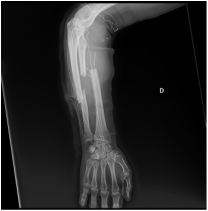
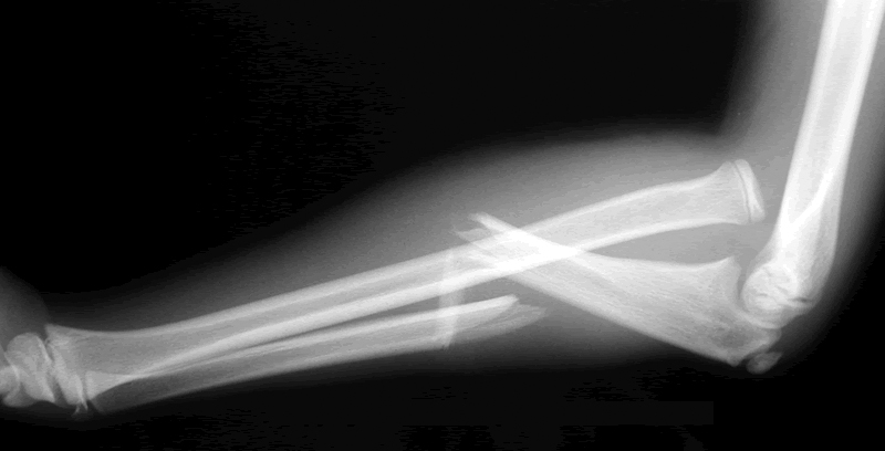
Lateral x-ray showing both radius and ulna fractures
Symptoms and Signs
  • Pain, swelling, deformity
  • Loss of function and movement
  • Assess elbow and wrist for associated injuries
  • Inform senior urgently if there is:
            - a possibility of compartment syndrome,
            - an open fracture
            - a neurovascular injury (unusual)
Initial Treatment
  • Analgesia
  • Above elbow backslab
  • Most will need fixation on unless isolated, undisplaced ulna fracture
Fixation
  • Plate fixation of both bones
  • Intramedullary nails in paediatric patients
Complications
  • Synostosis: 3-9%. Less with two surgical approaches
  • Infection
  • Compartment syndrome
  • Nonunion
  • Malunion
  • Re-fracture
  • Neurovascular injury
Monteggia fracture

Monteggia forearm fracture dislocation


  • Ulna # with radial head dislocation
  • More common in children
  • Admit for MUA or ORIF
Galeazzi Fracture

Galeazzi forearm fracture dislocation


  • Radial shaft fracture and dislocated distal radio-ulna joint (DRUJ)
  • Admit for ORIF of radius and reduction +/- stabilization of DRUJ
Powered by Create your own unique website with customizable templates.