JUNIORBONES
Orthopaedics and trauma for junior orthopaedic trainees and medical students
  • Home
    • About
  • Patient evaluation
    • History
    • Examination >
      • Shoulder examination
      • Hip examination
      • Knee examination
      • Foot & ankle examination
      • Spine examination
  • Trauma
    • Orthopaedic emergencies
    • Common trauma referrals >
      • Proximal humerus injuries >
        • Shoulder dislocation
        • Proximal humerus (shoulder) fractures
      • Upper-limb long bone fractures
      • Elbow injuries >
        • Adult elbow fractures
        • Paediatric supracondylar elbow fracture
      • Distal radius fractures
      • Spine trauma
      • Pelvic & acetabular fractures
      • Proximal femur fracture
      • Lower-limb long bone fractures
      • Knee injuries >
        • Fractures at the knee
        • Soft tissue knee injuries
      • Foot & ankle injuries >
        • Ankle fractures
        • Foot fractures
      • Peri-prosthetic fractures
      • Paediatric trauma
      • Septic arthritis >
        • Septic native joints
        • Septic joint replacement
  • Orthopaedic conditions
    • Osteoarthritis
    • Inflammatory & infective arthritis
    • Other joint disorders
    • Spine
    • Paediatric orthopaedics
    • Orthopaedic pathology
  • Cases blog
  • Trauma guidelines
  • Book downloads
  • Authors
  • Contact

Assessment and management of pelvic and acetabular trauma
by James Donaldson

Scenario: called to A&E to assess a 34 year old involved in a high speed road traffic accident.

History:

Age – often young, involved in high energy blunt trauma
Assess for other injuries

Time/date of injury

Past medical/surgical history

Last ate / drank
– timing of emergency surgery

Medication/drugs and allergies

Pelvic Trauma

Radiographs / imaging
  • Trauma series of x-rays (chest, AP pelvis and lateral cervical spine)
  • FAST scan (to rule out intra-abdominal bleeding)
  • CT scan only when haemodynamically stable
Symptoms and Signs
  • Pain, inability to weight bear
  • Assess lower limb: shortening, rotationally malaligned
  • ‘Balloting’ the pelvis has little value and can disrupt tamponade
  • Skin
            - Perineal lacerations
            - De-gloving
            - Flank haematoma
  • Uro-genital injury
            - Haematuria, scrotal bruising, blood at meatus
            - Do urethrogram prior to Foley. May need supra-pubic catheter
            - PR and PV mandatory - ?open #
  • Neurology:
            - Lumbosacral injury
            - Anal sphincter tone
Inform senior urgently
Advanced Trauma Life Support (ATLS)
  • Haemorrhage is the major source of death
  • Mortality 15-50%
  • Often associated injuries – chest, long bone, head, spine, abdomen, uro-genital
  • Resuscitation with blood, platelets and FFP (1:1:1)
  • Pelvic binder or sheet over greater trochanters, can combine with internal rotation of lower limbs
  • Sources of bleeding
            - Intra-abdominal
            - Intra-thoracic
            - Retroperitoneal
            - Extremity
            - Pelvic
                - Venous (80%)
                        - Post venous plexus
                        - Cancellous bone
                - Arterial (10-20%)
                        - Sup gluteal artery
                        - Int pudendal artery
                        - Obturator artery

Trauma team and seniors involved early
External fixator application


Angiography
  • To be done in theatre only, may be combined with laparotomy

  • May be helpful depending on resources
  • More useful for arterial bleeding
  • Perform after pelvis has been stabilised
Classification
Tile
    A) Stable
    B) Rotationally unstable
    C) Rotationally and vertically unstable
Young and Burgess
    AP compression type
    Lateral compression
    Vertical shear
BOAST guidelines:
  • Early pelvic binder and control of haemorrhage
  • PRC, platelets, FFP initially; if ongoing haemorrhage and pelvis is stabilized a laparotomy with pelvic packing +/- embolisation may be needed
  • Early CT scan once haemodynamically stable
  • High index of suspicion for associated injuries
  • Open pelvic fractures may require urgent bladder drainage and bowel diversion by specialists
  • Urethral injury diagnosed and managed appropriately
  • Definitive plan by pelvic surgeon within 5 days
  • Image transfer and referral to specialist centre within 24hrs
  • Specialist unit to follow the patient up

Acetabular trauma

Bimodal age distribution – young, high energy; elderly low energy

Radiographs
  • AP pelvis initially
  • May also need (ask specialist centre)
            - Judet views
            - Inlet and outlet views
            - +/- CT scan


acetabular fracture
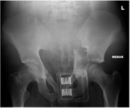
AP x-ray demonstrating Left acetabular fracture and pelvic binder insitu
Symptoms and Signs
  • Pain, swelling, inability to weight bear
  • Abnormal lower limb position / profile
  • 50% have an associated injury
  • 36% have an ipsilateral limb injury
  • Document neurovascular status
Management
  • In the high energy cases follow ATLS principles and management similar to pelvic fractures
  • A dislocated hip needs emergent reduction
  • Skin traction in A&E may be helpful
  • Inform senior urgently if high energy injury, haemodynamic instability, neurovascular compromise, open fracture or dislocation
  • Admit all cases for CT scan, analgesia and discussion
Letournel classification
  • Elementary fracture types (20%):
            - Anterior wall, anterior column, posterior wall, posterior column, transverse fracture
  • Associated fracture types (80%)
            - Involve 2 or more of the elementary types
Surgery
  • Non-surgical if minimally displaced or medically unfit
  • ORIF +/- total hip replacement if displaced
Complications
  • Secondary osteoarthritis
  • Heterotopic ossification
  • Femoral head avascular necrosis
  • DVT, PE
  • Nerve injury
  • Infection, bleeding, weakness, nerve injury  if surgery is performed
BOAST guidelines:
  • Reduce hip dislocations urgently. Document neurovascular status before and after the procedure. Apply skeletal traction. If irreducible or unstable urgent specialist transfer
  • CT scan within 24 hours of hip reduction and early referral / transfer of images
  • Reconstruction in a specialist unit within 5 days (no later than 10)
  • Chemical thromboprophylaxis within 48hrs of injury assuming no contraindications

Powered by Create your own unique website with customizable templates.