Infected joint replacements
by James Donaldson
Despite sophisticated prevention strategies infection rates of around 1% persist in elective joint arthroplasty and much higher rates following fixation for trauma.
Biomaterials and other foreign materials are inanimate and are thus susceptible to bacterial colonisation. After implantation implants are coated by the body with a layer of protein and platelets. If bacteria reach the implant prior to this coating by the host’s cells, bacterial adhesion may progresses to aggregation. In so doing these colonizing microorganisms develop and slowly grow in a structure known as a biofilm.
Existence within this biofilm represents a survival mechanism by which microbes resist external and internal environmental factors, such as antimicrobial agents and the host’s immune system.
History:
Age
Time/date of onset
Systemic symptoms - Pyrexia, night sweats, weight loss
Ability to ambulate and mobilise
When was the joint replacement carried out? Were there any post-operative problems? Antibiotic treatment post-op? Wound healing issues? Ongoing pain and swelling?
Recent illnesses or infection?
Gradual or sudden decline in function
Risk factors for infection – diabetes, immunosuppressed, drugs, elderly.
Past medical/surgical history
Fitness for surgery
Medication/drugs and allergies
Social history:
Can they cope at home?
Is the wound discharging, how much? What does it look like? Who is dressing it? Have they seen the GP? Any antibiotics started?
Clinical features:
Early post-operative infection
Late chronic infection
Haematogenous infection
Joint aspiration does not need to be done urgently. Usually this can be reserved until availability of aspiration in an operating theatre to guarentee sterile conditions.
Early post-operative infection
- Immediate post-operative period
- The wound may be erythematous, swollen, discharging and tender
- It may be difficult to differentiate between a superficial or deep infection (deep to fascia)
- Implant salvage remains possible at this stage
Late chronic infection
- Likely to originate at the time of surgery but a low virulence or inoculum delays the onset of symptoms
- Removal of the device is usually needed to eradicate the infection
Haematogenous infection
- Sudden, rapid deterioration in the function of an implant
- Most are some years down the line presenting with symptoms and signs similar to early post-operative infection
- It may be triggered by infection elsewhere, for example dental surgery, urinary sepsis or remote infection
- Implant salvage is possible if treatment is prompt
Joint aspiration does not need to be done urgently. Usually this can be reserved until availability of aspiration in an operating theatre to guarentee sterile conditions.
Investigations:
Consider:
Management:
Salvage procedures:
- Bloods: FBC, CRP, ESR, renal profile initially, blood cultures if patient septic
- Radiographs (likely to be normal but may show septic loosening in the late stages)
- Aspiration in theatre (70% sensitive) - send for microscopy & culture/sensitivity
Consider:
- Nuclear imaging - bone scan (will be ‘hot’ for 2 years post surgery) or labeled white cell scan
Management:
- For acute (post-operative or haematogenous infection)
- Extensive and meticulous debridement with retention of the prosthesis
- Continue antibiotics until wound & inflammatory markers settle (usually 6 weeks minimum)
-
Needs to be done within 6 weeks to prevent biofilm formation ~90% success rate
- Revision surgery for chronic infections - 1 or 2 stage procedure with long-term antibitoics (usually 6 weeks minimum)
Salvage procedures:
- Arthrodesis – usually performed in native joint infection - largely historical procedure now
- Excision arthroplasty – often reserved for the moribund or where the joint is unreconstructable
- Amputation / disarticulation – when all else has failed
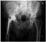
First stage revision with cement spacer in situ
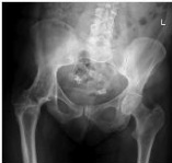
Arthrodesis (fusion) of native joint for previous infection
|
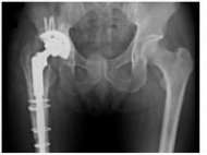
Second stage revision total hip replacement
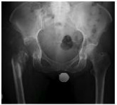
Excision arthroplasty (also known as Girdlestone procedure) following infected total hip replacement
|
