Assessment and management of fractures around the elbow in adults
by James Donaldson
SCENARIO: you are called to the Emergency department to assess a 20 year old who fell from a height onto his elbow. No other injuries.
HISTORY:
Age
Occupation – useful for deciding treatment
Handedness
Time/date of injury
Past medical/surgical history
Medication/drugs and allergies
Last ate/drank (for timing of emergency surgery if needed)
Immediate assessment:
Exclude all other injuries
Examine affected limb - look for obvious deformity and determine if open or closed injury.
Examine radial/ulna/median nerves both motor and sensation. Palpate for a radial & ulna pulses. Assess capillary refill.
Investigations:
X-Ray of affected limb - true AP and lateral of elbow with images of joint above and below
X-Ray of affected limb - true AP and lateral of elbow with images of joint above and below
- Assess bony contours and presence of fractures / dislocations
- Anterior fat pad sign is pathopneumonic for an intra-articular injury
- The anterior humeral line should intersect the anterior third of the capitellum
- The radial head and capitellum should line up on both views
|
Injury
Olecranon fracture Radial Head Fractures Elbow dislocation Distal humerus fractures |
Mechanism
Direct violence – fall on point of elbow or direct blow Indirect - fall on outstretched hand with elbow in flexion and contraction of triceps Fall on outstretched hand, valgus loading or direct trauma Fall onto extended elbow High energy trauma or fall in adults |
Signs & symptoms
|
|
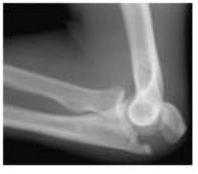
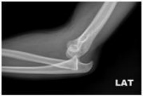
Olecranon fracture
Radial head fracture
|
|
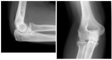
Elbow dislocation
|
|
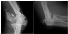
Distal humerus fracture
Call senior if: Neurovascular compromise or open injury |
