Assessment and management of acute soft tissue knee injuries
by Francois Tudor
Scenario: called to A&E to assess a 20 year old man who hurt his knee whilst playing soccer today. No other injuries.
History
- Age
- Occupation – useful for deciding treatment
- Sports level and aspirations – very important for deciding definitive treatment plan
- Time/date of injury
- Mechanism
- Immediate problem – pain/swelling/knee collapsing
- Able to continue playing – can indicate severity of injury (but not always - beware)
- Immediate treatment – ice, strapping, physio
- Problem now – particularly if not an acute injury – pain, swelling, instability, locking (unable to get knee fully straight)
- Past medical/surgical history
- Previous knee problems/surgery
- Medication/drugs and allergies
|
Mechanism
Contact to knee: Fracture Posterior cruciate ligament (PCL) injury Multiple ligament disruption Knee dislocation Contact from lateral side of knee: Medial collateral ligament (MCL) injury ± other ligament injury meniscal tear Non-contact: Anterior cruciate ligament (ACL) injury Meniscal tear Twist on planted leg: Anterior cruciate ligament (ACL) injury Meniscal tear Patella dislocation |
Impact on anterior knee/shin: Posterior cruciate ligament (PCL) injury Patella fracture Impact to knee cap: Patella fracture Patella dislocation Paediatric knee injury: Ligament avulsion Growth plate injury Hyperflexion/resisted extension: Quadraceps/patella tendon rupture (often >30 years old) |
Examination
Antalgic gait or non-weight bearing - Fracture, meniscal tear, ligament injury
Unable to straight-leg raise - Quadriceps/patella tendon rupture or patella fracture
Large effusion - Intra-articular fracture, ACL rupture, meniscal tear
Reduced range of motion (Normal -2 to 130 degrees) - Fracture, meniscal tear, ligament injury, isolated effusion
Block of full extension - Locked bucket-handle tear of meniscus
Joint line tenderness - fracture, meniscus tear
Anterior drawer/Lachman +ve - ACL rupture, (possible false +ve with PCL injury)
Opening to valgus stress - MCL injury
Opening to varus stress - LCL injury
Investigation
ALWAYS send for knee x-rays, even when suspicion is of isolated soft-tissue injury.
Request – AP (weight-bearing if possible), lateral and skyline x-rays of knee.
Look for:
- Fracture – tibial plateau, distal femur, minor fractures (Segond fracture of lateral plateau seen with ACL rupture)
- Patella fracture/dislocation
- ACL bony avulsion from tibia
ALWAYS send for knee x-rays, even when suspicion is of isolated soft-tissue injury.
Request – AP (weight-bearing if possible), lateral and skyline x-rays of knee.
Look for:
- Lateral view:
- Fracture – tibial plateau, distal femur, minor fractures (Segond fracture of lateral plateau seen with ACL rupture)
- Patella fracture/dislocation
- AP view:
- ACL bony avulsion from tibia
|
Management
Knee dislocation (with or without fracture) Assess neuro-vascular status – IF ANY DOUBT, call vascular surgeons Call senior urgently Reduce and backslab/brace (may require theatre) Admit for further investigation/treatment Monitor for vascular compromise Fracture (tibial plateau, patella, distal femur), growth plate injury or avulsion Analgesia Assess neuro-vascular status Assess for open fracture Apply backslab Elevate and non-weight bearing Admit to ward Monitor for compartment syndrome Discuss with senior Suspected quadriceps/patella tendon rupture Analgesia Backslab/brace if painful Admit Investigate with Ultrasound Locked bucket handle meniscus tear Analgesia Non-weight bearing (brace if very painful) Urgent investigation with MRI and review <1 week Discuss with senior ACL/PCL injury Analgesia Crutches and brace as necessary Ice and gentle range of motion Review in clinic 1 week Investigate with MRI as outpatient ACL injury with valgus/varus opening (suspected MCL/LCL injury) Analgesia Crutches and brace Ice and elevation Review clinic < 1 week Meniscus tear (with full ROM) Analgesia Crutches non-weight bearing Review in clinic 1 week Investigate with MRI as outpatient Any patients with high energy injury or severe mechanism but with a seemingly innocuous injury should be reviewed by senior orthopaedic doctor before discharge. |
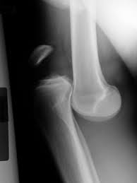
Lateral x-ray knee demonstrating knee dislocation. THIS IS AN EMERGENCY
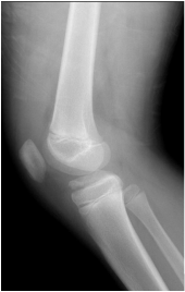
Lateral x-ray of knee of skeletally immature patient demonstrating displaced avulsion fracture of the ACL origin
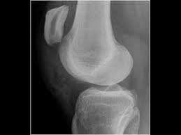
Lateral x-ray knee demonstrating high riding patella due to patellar tendon rupture
|
